Resting state fNIRS signal decomposition
Functional near infrared spectroscopy (fNIRS) is a non-invasive brain imaging technique that is based on the spectroscopic measurement of the optical absorption of cerebral blood. FNIRS can be used to characterize spontaneous resting brain activity. Both oxyhemoglobin (Oxy-Hb) and deoxyhemoglobin (deoxy-Hb) concentration changes can be estimated from the modified Beer-Lambert law using absorption measurements taken at multiple wavelengths of light within the optical window (650-850nm). FNIRS imaging is performed using a set of spatially arranged optical sources and detectors (optodes), which are often coupled through fiber optics between a head cap worn by the participant and the fNIRS instrument. A potential limitation of fNIRS is contamination from unwanted physiological noise related to autonomic blood pressure, respiratory signals, and cardiac pulsation. The sensitivity of fNIRS measurements falls off exponentially with depth to reach a penetration of approximately 5-10mm into the cortex of the brain. This results in a over-sensitivity to superficial physiological noise in the scalp, especially due to the skin flow. Contamination by systemic physiology poses several challenges to the analysis and interpretation of fNIRS neuroimaging data. The functional connectivity within different cerebral regions can be highly affected by physiological noise. In the presence of systemic physiology, it is difficult to determine the portion of the fNIRS signals attributed to oscillations arising from neural connectivity in the brain. The characterization of slow resting-state oscillations in brain activity for connectivity analysis poses additional challenges in fNIRS due to the contamination of additional low-frequency vascular changes with no direct coupling to neuronal activities. These vascular changes are regional and may be generated by the auto-regulation of regional cerebral blood flow.
In a study published in 2016 (Aarabi and Huppert, 2016), we presented a single-channel decomposition method (Fig. 1) called single-channel ICA (scICA) to characterize the contributions of low-frequency oscillations, respiration, and cardiac pulsation in oxyhemoglobin, deoxyhemoglobin, and total-hemoglobin signals. The scICA method, as a subspace approach, was used to decompose these time series into their underlying sources, which were then used to estimate the contribution of the constituent components. We applied the scICA method to the resting-state fNIRS data from a dataset of 17 human subjects. The contribution of various sources of physiological noise was then estimated for different cerebral regions across the head. We evaluated the results at single-subject and group levels.
The scICA method comprises three main stages, preprocessing, pre-decomposition, and single-channel decomposition. In the preprocessing step, the fNIRS signals are first band-pass filtered. Noisy channels are then rejected. Finally, motion artifacts are removed using the spatial ICA. The clean fNIRS signals of all channels are decomposed using the scICA method. An automatic procedure is used to estimate the optimal number of independent components (sources) for each channel based on the identification of the frequency bands associated with low-frequency oscillations, respiration, and cardiac activity as well as those related to cardio-respiratory interactions. Finally, independent components associated with each frequency band are identified and projected back onto the measurement space to reconstruct the multichannel fNIRS signals corresponding to that frequency band.
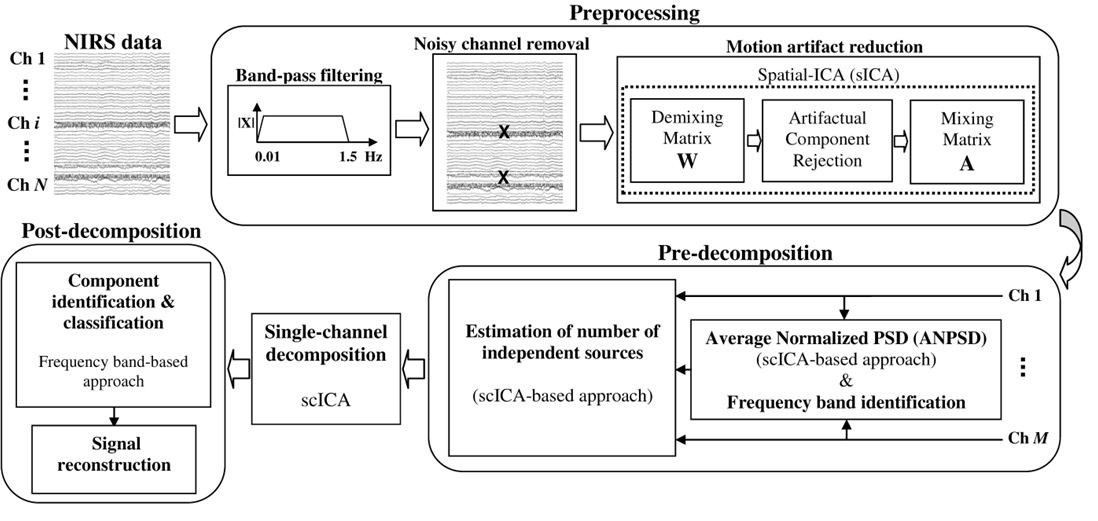
Figure 1. Schematic of the single-channel decomposition process.
Our results indicated that low-frequency oscillations account for 40-55% of total power over the whole spectrum of both the oxygenated and deoxygenated hemoglobin signals. Respiration and its harmonics accounted for 10-30% of the power, and cardiac pulsations and cardio-respiratory components accounted for 10-30%. Our results also showed a major contribution of the brain autoregulatory activity and local brain vascular autoregulation, (33-38% for very low-frequency components, <0.08Hz ) and (4-9% for low-frequency components, 0.08-0.15Hz) to the resting state deoxy-Hb, Oxy-Hb, and total-Hb signals. Our approach is applicable to both single and multiple-channel fNIRS signals. It also allows functionally distinct sources of noise with disjoint spectral support to be separated from obscuring systemic physiology (Aarabi and Huppert, 2016).


References
Aarabi, A., Huppert, T.J., 2016. Characterization of the relative contributions from systemic physiological noise to whole-brain resting-state functional near-infrared spectroscopy data using single-channel independent component analysis. Neurophotonics 3, 025004.